
Understanding What is MRI Scan?
Magnetic Resonance Imaging (MRI) is a revolutionary non-invasive imaging technology that plays a vital role in modern medicine. This sophisticated medical tool enables healthcare professionals to create detailed three-dimensional anatomical images, aiding in disease detection, diagnosis, and treatment monitoring. MRI is particularly valuable for visualizing soft tissues and comes with various applications and considerations.
How MRI Works
- At the heart of MRI technology are powerful magnets that generate a strong magnetic field within the machine. This magnetic field causes the protons found in the water molecules within living tissues to align themselves with the field’s direction. When a radio frequency current is applied, it excites these protons, causing them to temporarily deviate from their alignment.
- As the radio frequency field is turned off, the protons gradually realign with the magnetic field. During this realignment process, they emit energy, which the MRI sensors detect.
- The time taken for protons to realign, as well as the energy released, varies depending on the specific tissue characteristics. Physicians can differentiate between different types of tissues based on these magnetic properties, creating detailed images.
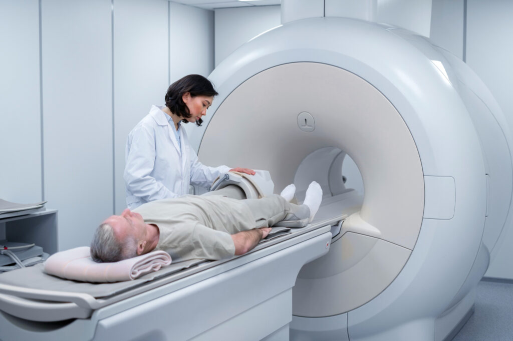
- To obtain an MRI image, a patient is positioned inside a large magnet and must remain perfectly still to prevent image blurring. In some cases, contrast agents containing Gadolinium may be administered intravenously to expedite the realignment of protons, resulting in a brighter image.
Applications of MRI
MRI is highly effective at imaging non-bony structures and soft tissues within the body, setting it apart from other imaging methods like X-rays and CT scans, which use ionizing radiation. Some key applications of MRI include:
- Neuroimaging: MRI is exceptionally well-suited for examining the brain, spinal cord, and nerves. It can distinguish between white and gray matter and is crucial in diagnosing conditions such as aneurysms and tumors.
- Musculoskeletal Imaging: MRI excels at visualizing muscles, ligaments, tendons, and joint injuries, making it a valuable tool in diagnosing orthopedic issues, especially knee and shoulder injuries.
- Functional MRI (fMRI): This specialized MRI technique is used to observe brain structures and their activation patterns during cognitive tasks. It contributes to our understanding of brain organization and neurological conditions.
- Abdominal and Pelvic Imaging: MRI is used to examine organs in the abdomen and pelvis, aiding in the detection and diagnosis of various conditions, including tumors and inflammatory diseases.
Risks and Considerations
While MRI does not expose patients to ionizing radiation like X-rays or CT scans, it does involve a powerful magnetic field. Several important considerations include:
- Implants: Patients with certain metal implants, such as pacemakers, cochlear implants, or certain types of neurostimulators, should not undergo MRI due to the risk of interference with these devices.
- Noise: MRI machines can be noisy, with sounds reaching up to 120 decibels. Special ear protection may be required.
- Nerve Stimulation: Rapidly changing magnetic fields during an MRI can cause a twitching sensation.
- Contrast Agents: Patients with severe renal failure may be at risk of a rare condition called nephrogenic systemic fibrosis when exposed to certain gadolinium-based contrast agents.
- Pregnancy: While there’s no conclusive evidence of harm to the fetus, MRI scans are generally avoided during the first trimester of pregnancy.
- Claustrophobia: Patients with claustrophobia might find MRI machines challenging to tolerate. Open MRI machines, which are less confining are available as an alternative.
In summary, MRI is a powerful and versatile medical imaging tool that provides invaluable insights into the human body. Understanding its principles, applications, and associated risks is essential for both healthcare professionals and patients alike.